Nucleocytoplasmic transport regulated by a phase-separated compartment

In eukaryotic cells, chromosomes are sequestered in a membrane-bound structure, the nucleus. This compartmentalization allows for complex and specialized regulation, but poses the challenge of exchanges between the nucleus and the cytoplasm. These occur through large multiprotein assemblies spanning the nuclear envelope, called Nuclear Pore Complexes (NPCs). Molecular transport through NPCs determines the cellular location of many macromolecules, for example transcription factors, and is therefore fundamental for the regulation of gene expression, cell division, and other critical cellular functions.

NPCs are equipped with a permeability barrier to control nucleocytoplasmic fluxes. Their central channel is densely packed with intrinsically disordered proteins. In vitro studies have revealed that these disordered proteins can undergo liquid-liquid phase separation (LLPS) into droplets that mimic the behavior of the NPC permeability barrier. This suggests a potential role for LLPS in the selectivity of nucleocytoplasmic transport.

This visual resource aims at explaining the mechanisms of nucleocytoplasmic transport and highlighting its crucial regulatory role in cellular activity. All animations can be downloaded by right-clicking and saving the corresponding file.

It was designed and implemented by Margot Riggi, under the supervision of Janet Iwasa and in collaboration with Ofer Rog (University of Utah). This work was funded through NSF grant 2219605.

We recommend visiting this webpage on a computer.

1. Overview of nucleocytoplasmic transport
1.1. Introduction to the key players
1.2. Animated summary of nuclear import and export

2. How LLPS-like mechanisms contribute to the NPC permeability barrier

3. A concrete example: nucleocytoplasmic transport plays a key role in the regulation of NF-kB signalling

1. Overview of nucleocytoplasmic transport

1.1. Introduction to the key players

5 µm Cross-section Mitochondria Golgi apparatus Endoplasmic Reticulum & Nuclear Envelope Nucleus Nuclear Pore Complexes Mitochondria
Chromatin Lamins mRNA NuclearEnvelope Ribosome Microtubule Actin Nucleus -cytoskeletonanchor Nuclear Pore Complex 30 nm 10 µm

The nuclear envelope does not create the same topological barrier as most other membrane-bound organelles.
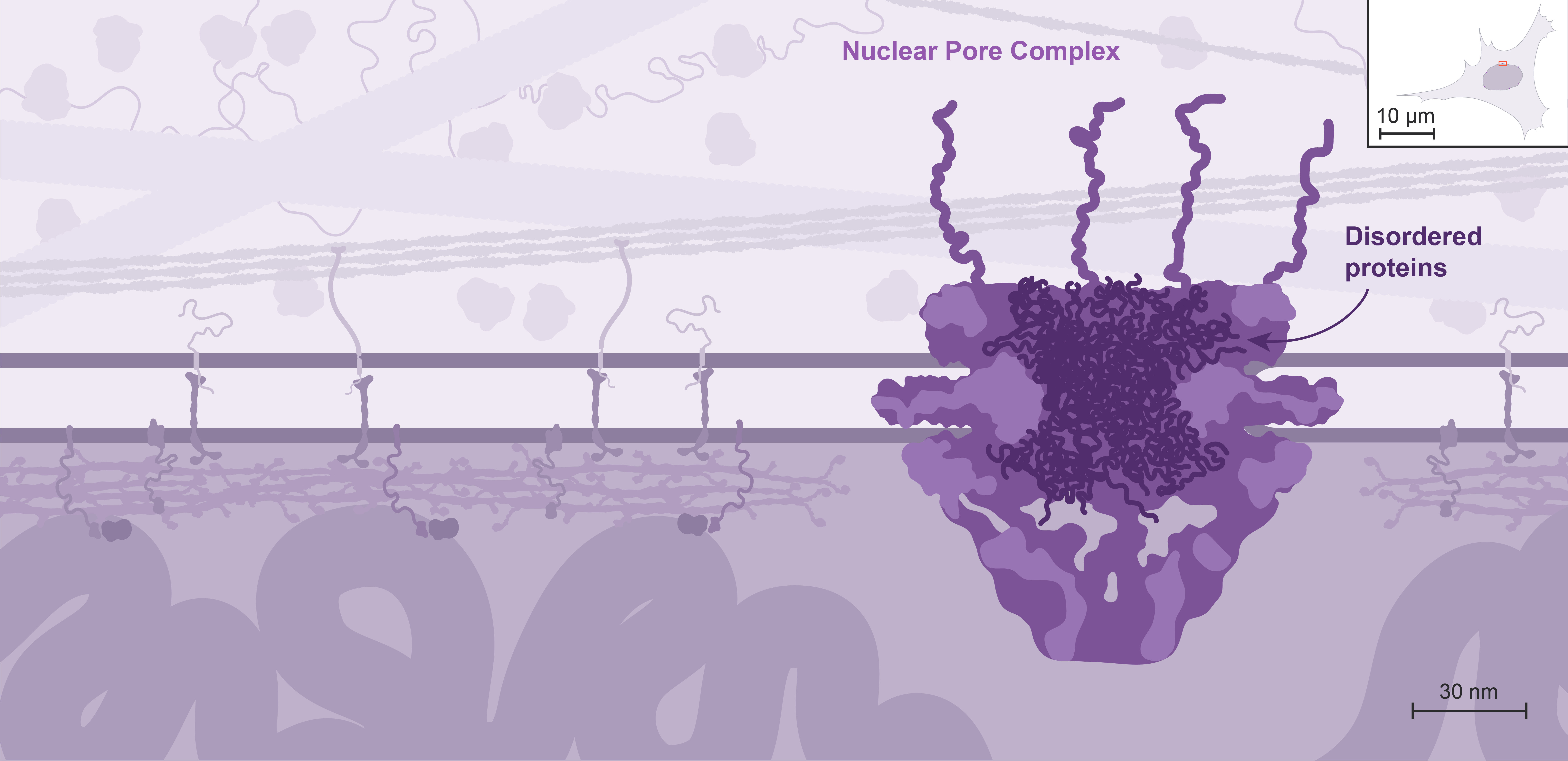
Instead, exchanges between the nucleus and the cytoplasm take place through large multiprotein channels called Nuclear Pore Complexes, or NPCs.
The nuclear envelope of a typical mammalian cell contains between 3000–4000 NPCs.

Mouse over the cell to explore the various organelles.

The nuclear envelope consists of two layers of membrane, enclosing a lumen, and is continuous with the endoplasmic reticulum.
The inner and outer nuclear membranes connect at the sites of the NPCs.

Mouse over the scene to explore all of its elements.

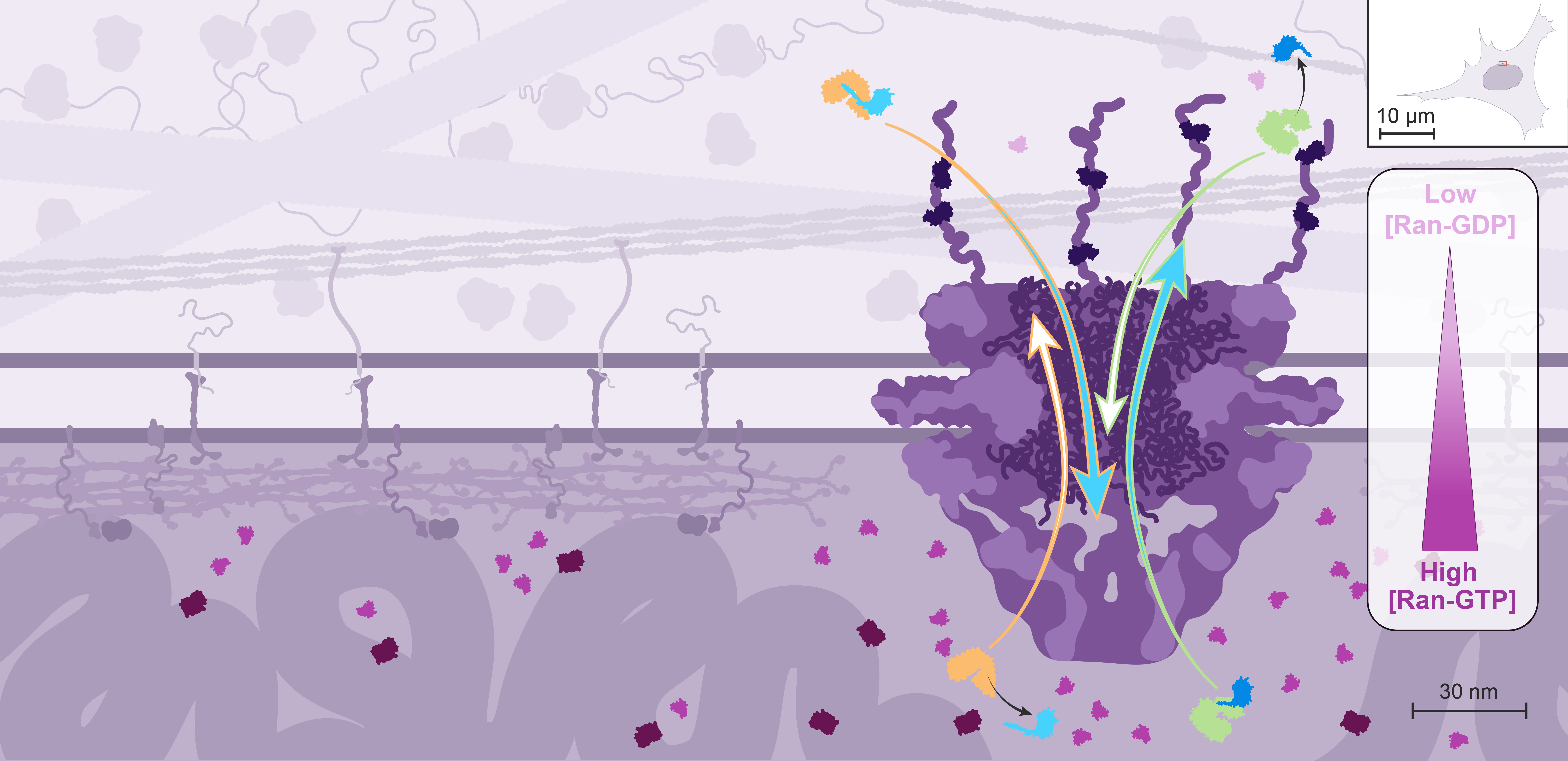
NPCs allow free passage of small molecules through diffusion, but regulate the transit of macromolecules as disordered proteins located within the central channel form a sieve that most macromolecules can’t cross by themselves.

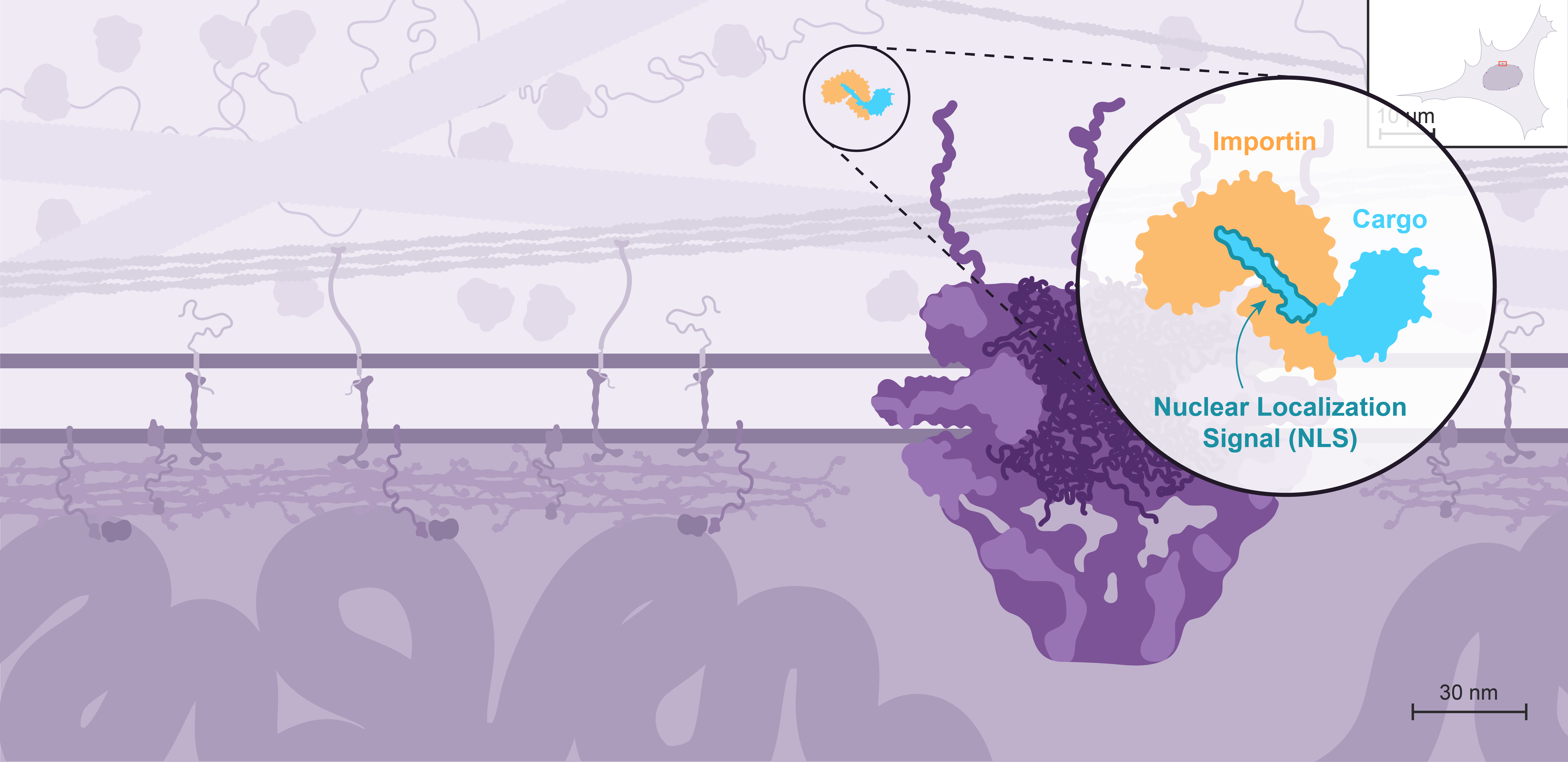
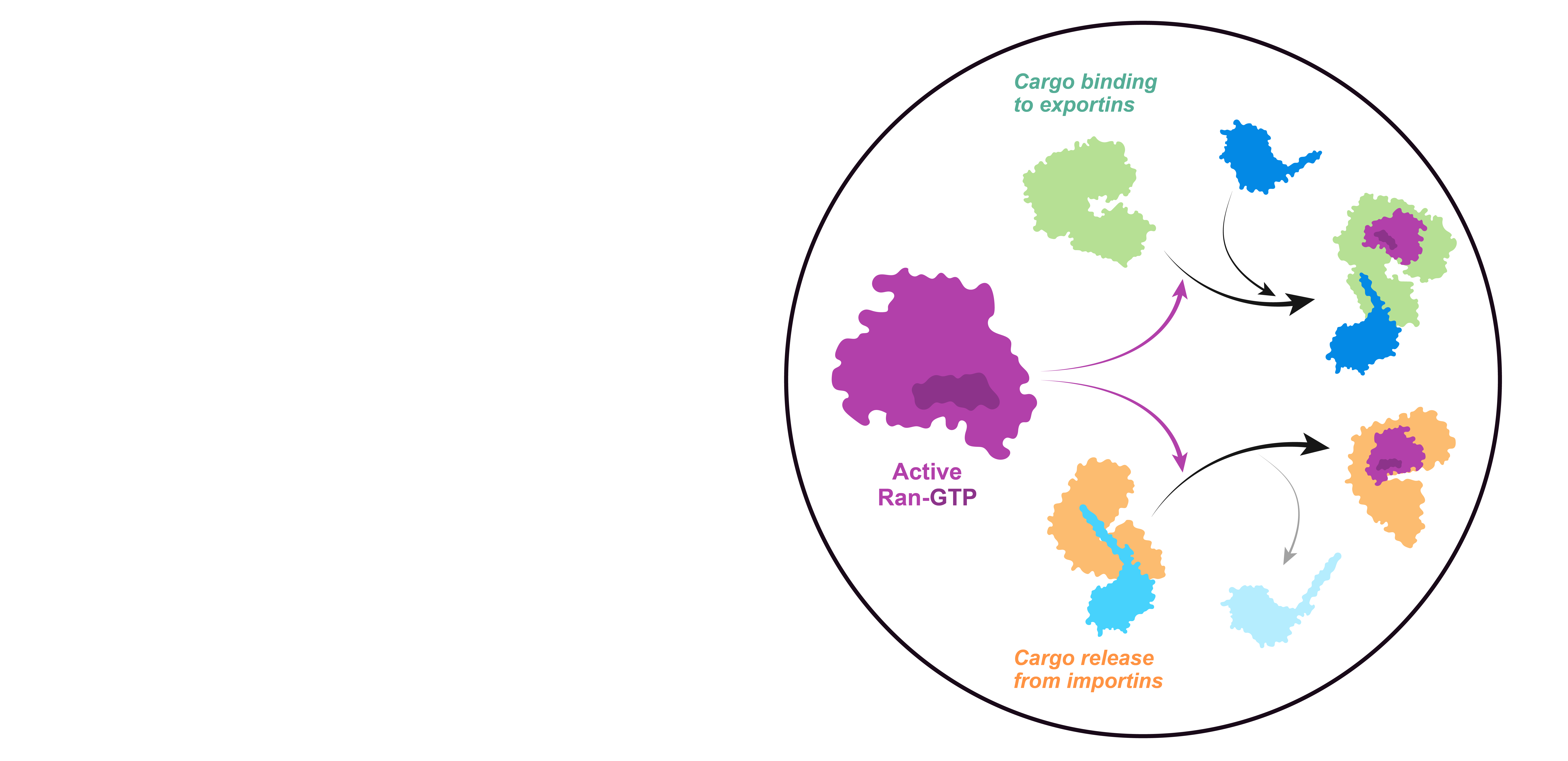
Nuclear import is mediated by transport receptors called importins, which recognize proteins containing a Nuclear Localization Signal, or NLS.
All the nuclear proteins - including structural organizers of the genome, transcription factors, and enzymes acting on DNA - are made in the cytoplasm and therefore need to get imported.

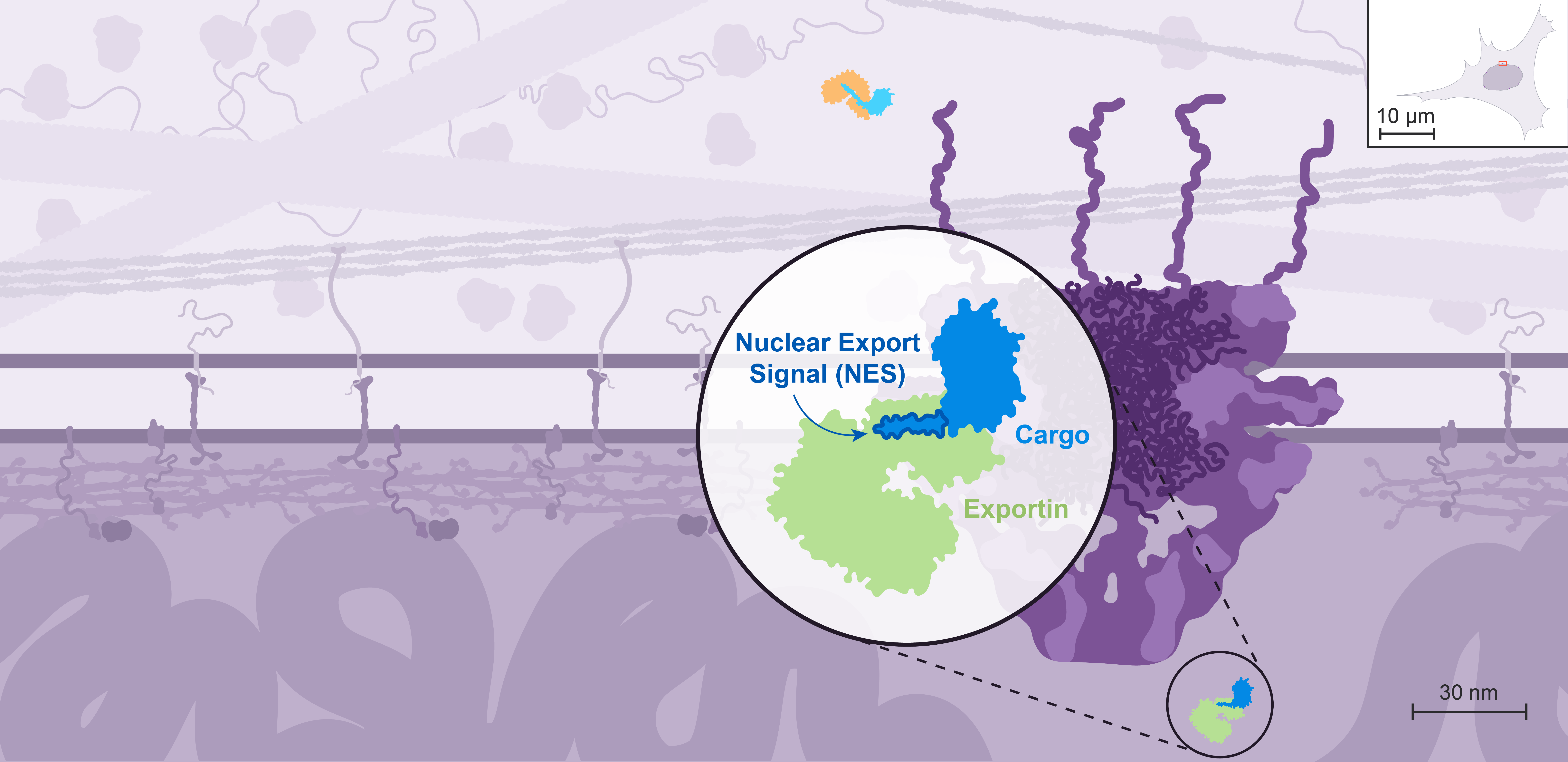
Nuclear export is mediated by different transport receptors called exportins, which recognize proteins containing a Nuclear Export Signal, or NES.
mRNA and ribosomes are examples of some of the elements that need to get exported.

Importins and exportins weakly interact with the disordered proteins in the central channel. This facilitates the passage of these transport receptors and their cargo through the NPC. Even giant cargo, such as ribosomes, can be transported through the NPC in only a few milliseconds.
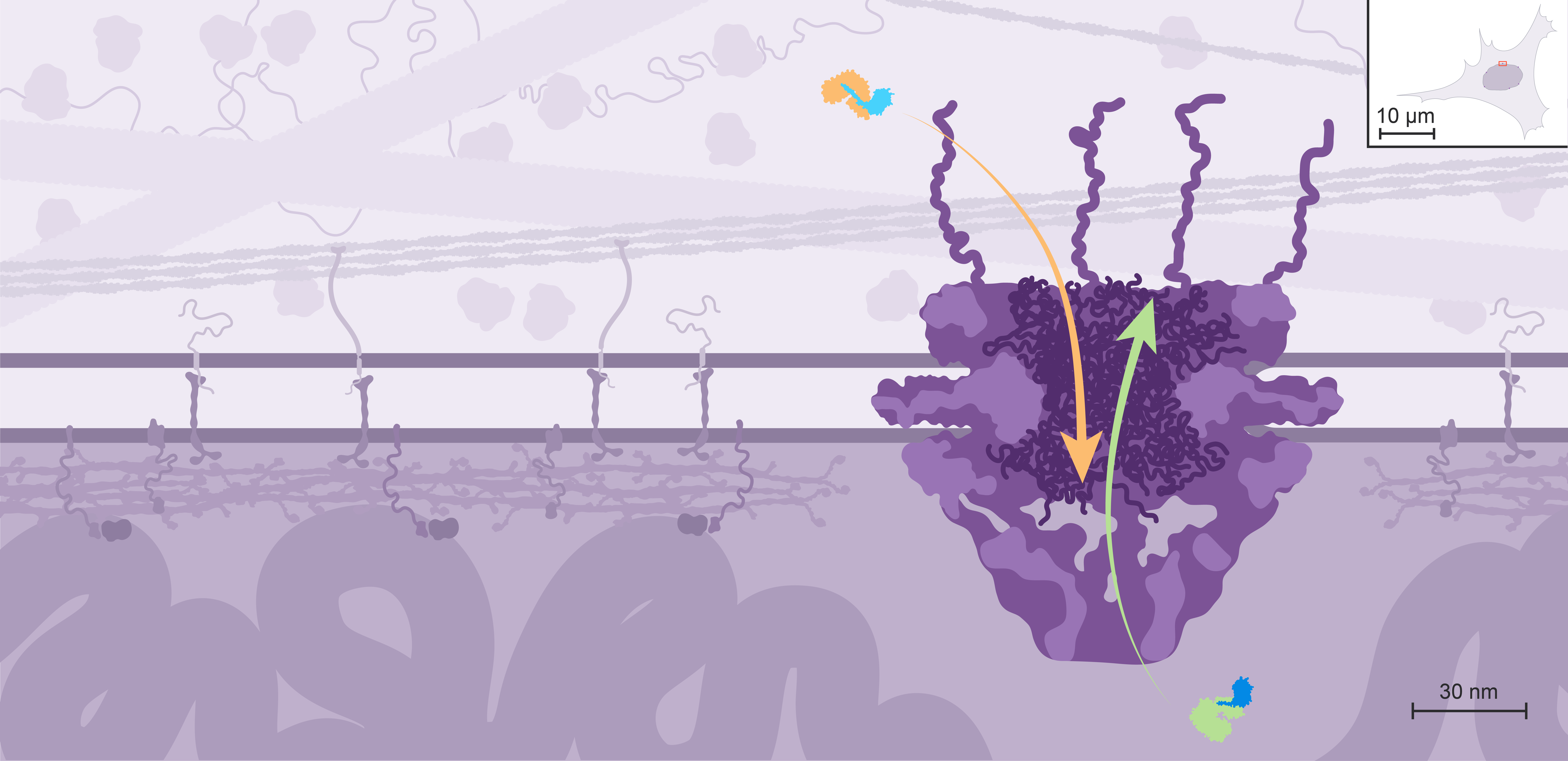
However, these random events cannot explain the directionality of nucleocytoplasmic transport, which can efficiently concentrate or exclude certain proteins. In this case, directional transport depends on the asymmetric distribution of the small GTPase Ran rather than on protein pumps.

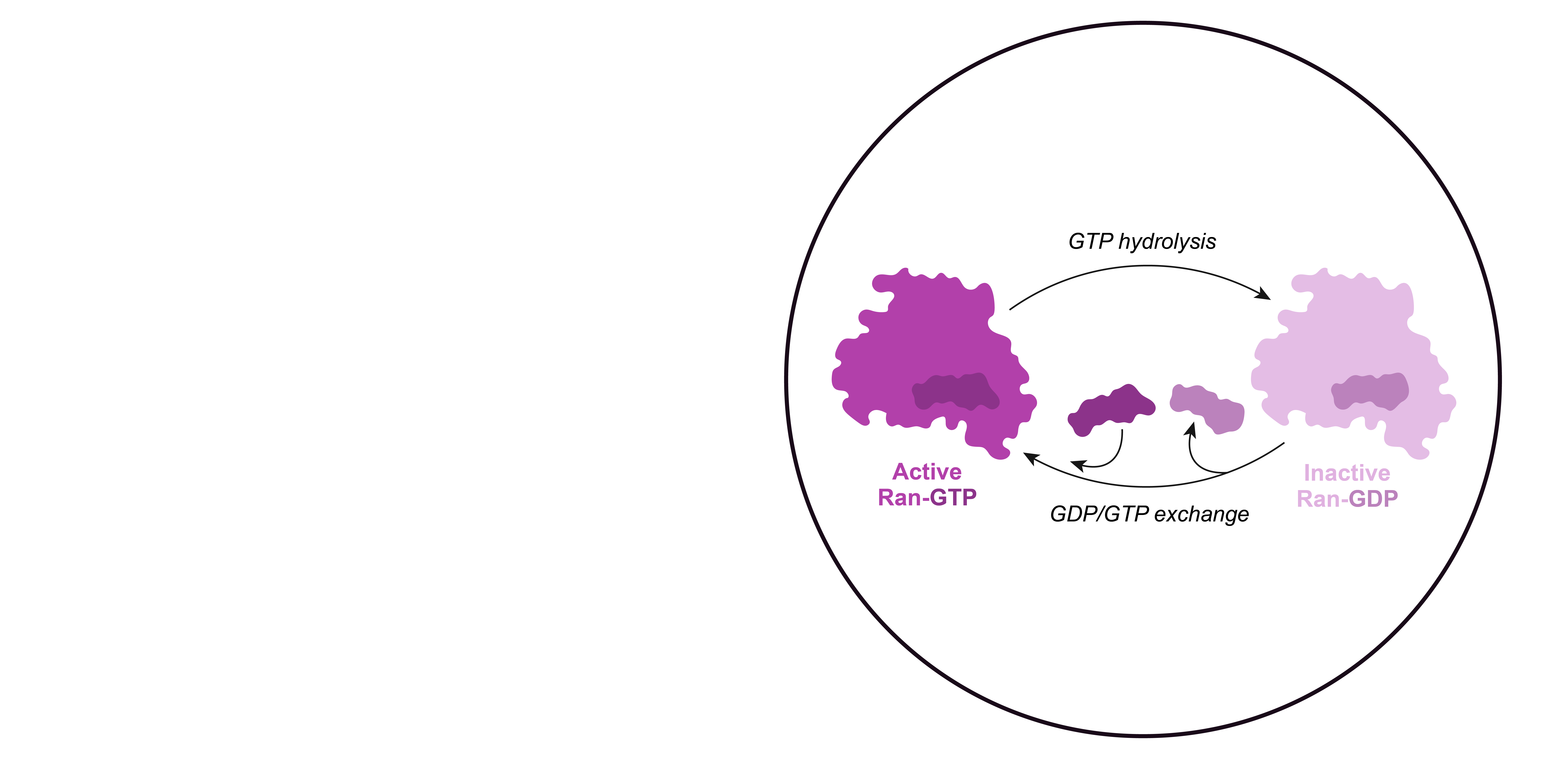
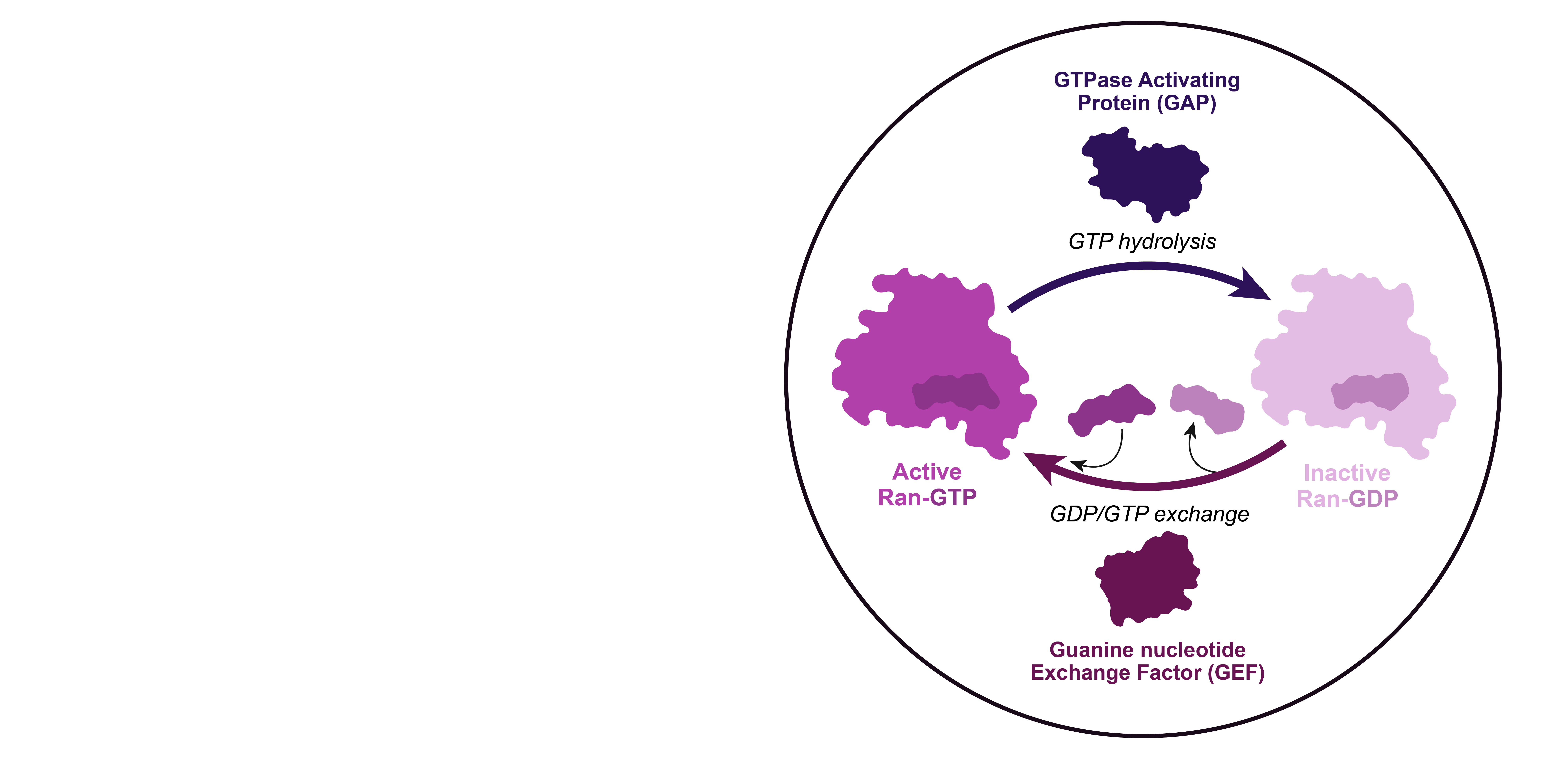
Small GTPases like Ran are active when bound to GTP, and inactivate upon GTP hydrolysis. The GDP can be then replaced by another GTP.

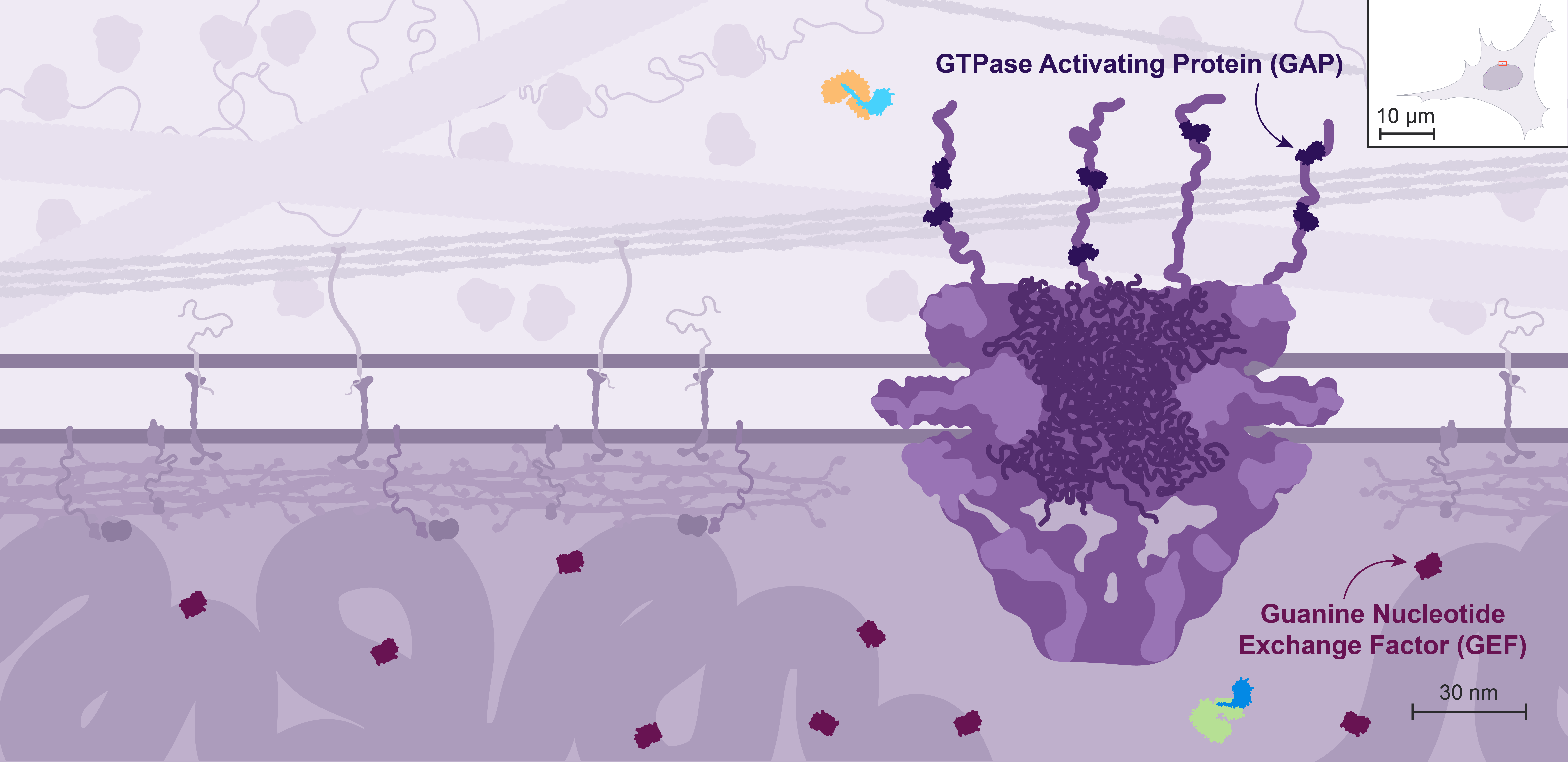
GTP hydrolysis by Ran is enhanced by GTPase Activating Proteins, or GAPs, while GTP exchange is promoted by Guanine nucleotide Exchange Factors, or GEFs.

Only the GTP-bound form of Ran can interact with high affinity with both importins and exportins, but with opposite effects on cargo binding: it competes with cargos for importin binding but promotes their binding to exportins.

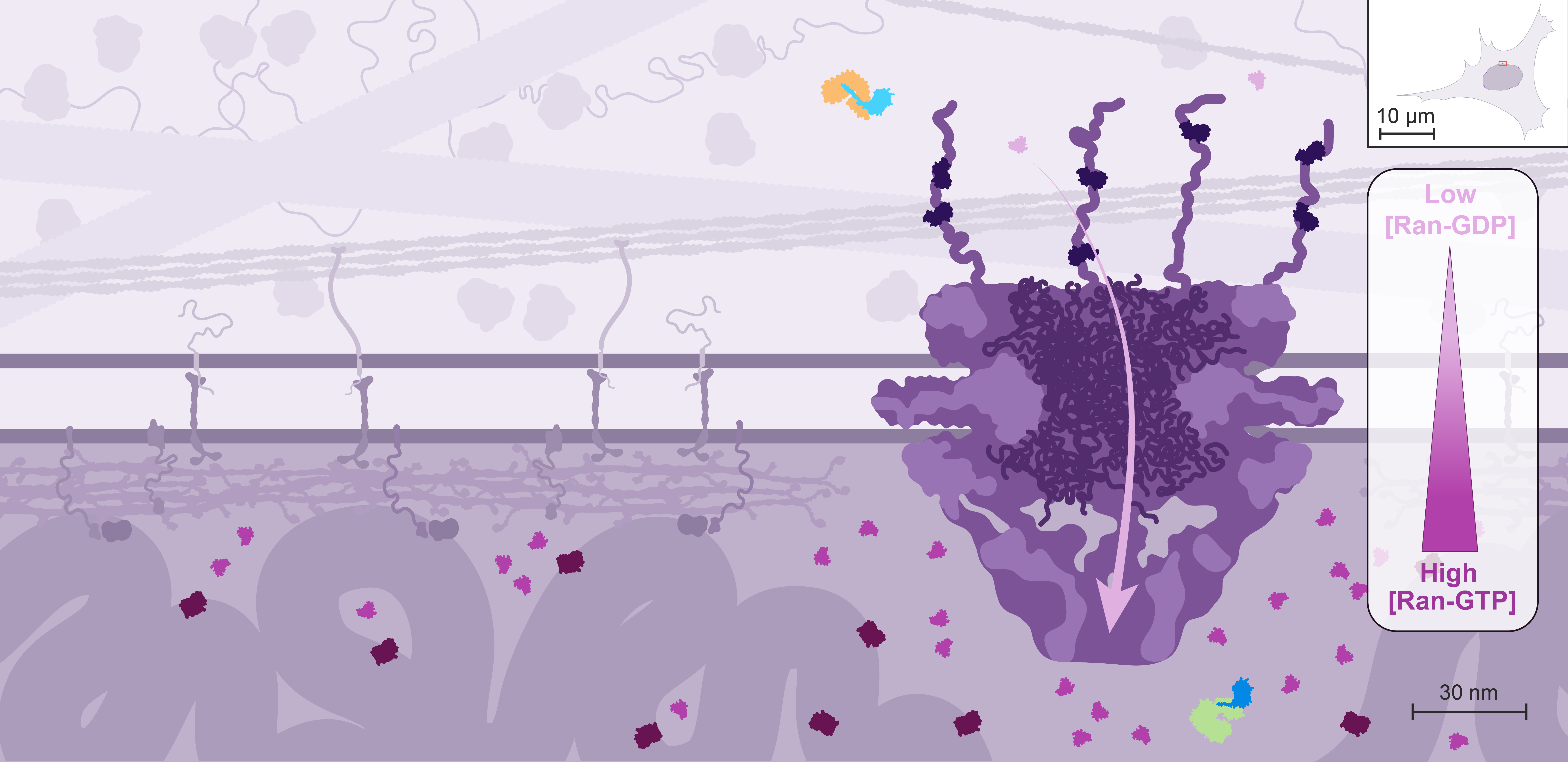
In the cell, the localization of Ran GEFs and GAPs is compartmentalized: the GEFs localize to the nucleus through association with the chromatin, while the GAPs are localized to the cytoplasmic side through interactions with components of the NPC.

As a result, Ran is predominantly loaded with GTP in the nucleus, and rapidly converted into the GDP-bound state in the cytoplasm. Ran-GDP is small enough to diffuse back passively into the nucleus, but its transport can also be facilitated by its own importin.
It is estimated that Ran-GTP is 200-fold enriched in the nucleus.

This Ran gradient controls the direction of nuclear import and export.
1.2. Animated summary of nuclear import and export

NUCLEAR IMPORT

NUCLEAR EXPORT

2. How LLPS-like mechanisms contribute to the NPC permeability barrier

The features of nuclear import and export have been well described, including the selective formation and dissociation of various complexes of cargo, as well as the roles of importins/exportins and Ran. However, the mechanism allowing the unique selectivity of the inner channel of the NPC remained unknown.
Such mechanism should enable size-based permeability on small scales, and size-independent but importin/exportin-dependent on larger scales. Most molecular mechanisms to explain such selectivity rely on intricately positioned protein interfaces to achieve tight regulation and specificity, but in the case of the NPC, an alternative hypothesis emerged: that these properties are the consequence of the properties of a dedicated material present in the inner channel.

The original observation leading to this understanding was the observation that proteins in the NPC central channel contain short repeated clusters of hydrophobic amino acids such as phenylalanines (called FG-repeats and corresponding to the “stickers” of the LLPS sticker/spacer model) separated by hydrophilic sequences (corresponding to the “spacers” of the model). Click here for more details about the sticker/spacer model.

Facilitated nucleocytoplasmic transport involves interactions of nuclear transport receptors with these FG repeats, which suggests that they might contribute to the selectivity of nucleocytoplasmic transport.

The following animations explain how the NPC disordered proteins, also called FG-nucleoporins or FG-Nups, form a functional permeability barrier.

2.1. FG-repeat domains of NPC disordered proteins form hydrogels in vitro

A highly concentrated solution of FG-repeat domain can form solid hydrogels in vitro. However, a similar solution remains liquid when all the hydrophobic phenylalanines are mutated into serines.
This experiment by Frey & al (Science, 2006) demonstrates that inter-repeat interactions between phenylalanines are responsible for the ability of the WT FG-repeat domain to form hydrogels.
The formation of this novel material hinted that the biophysical properties of the NPC inner channel might hold the key to the mechanism that determines permeability.

2.2. FG-Nups form a selective permeability barrier through transient interactions

The previous observation supports a model in which the NPC permeability barrier forms a sieve-like structure through hydrophobic interactions between the FG-repeats of disordered nucleoporins, while the mesh size sets the size limit for passive exclusion of molecules: it allows passage of small molecules but block fluxes of free macromolecules larger than 30 kDa.
According to this model, nuclear transport receptors can overcome this size restriction because their binding to hydrophobic FG-repeats competes with the inter-repeat interactions and transiently opens adjacent meshes of the sieve.
Hydrogels formed by highly concentrated FG-repeat domains have also been shown to reproduce these permeability properties in vitro (Frey & Görlich, Cell, 2007).

2.3. In vitro intra-droplet dynamics of FG-Nups and nuclear transport receptors reproduces their in vivo behavior

Fluorescence Recovery After Photobleaching (FRAP) is a method used to determine the diffusion kinetics of fluorescently-labeled molecules. A small region of the field of view is submitted to high intensity illumination, or “photobleached”, which irreversibly eliminates the fluorescence of the fluorophores present in this area, while their other biological properties remain unaffected. Because of Brownian motion, the molecules labeled with still-fluorescent fluorophores diffuse throughout the sample, including in the bleached region. The faster the molecular diffusion is, the faster this fluorescence recovery will occur. Click here for more details about FRAP.

This FRAP experiment by Schmidt & Görlich (Elife, 2015) reveals the low mobility of phase-separated FG-Nups, tagged in red, while nuclear transport receptors (here the importin NTF2, tagged in green) show a very high mobility within these FG-Nup droplets. This behavior is reminiscent of nuclear transport receptors diffusing through the central channel of NPCs.

2.4. In vitro experiments with FG-Nup droplets recapitulate in vivo permeability

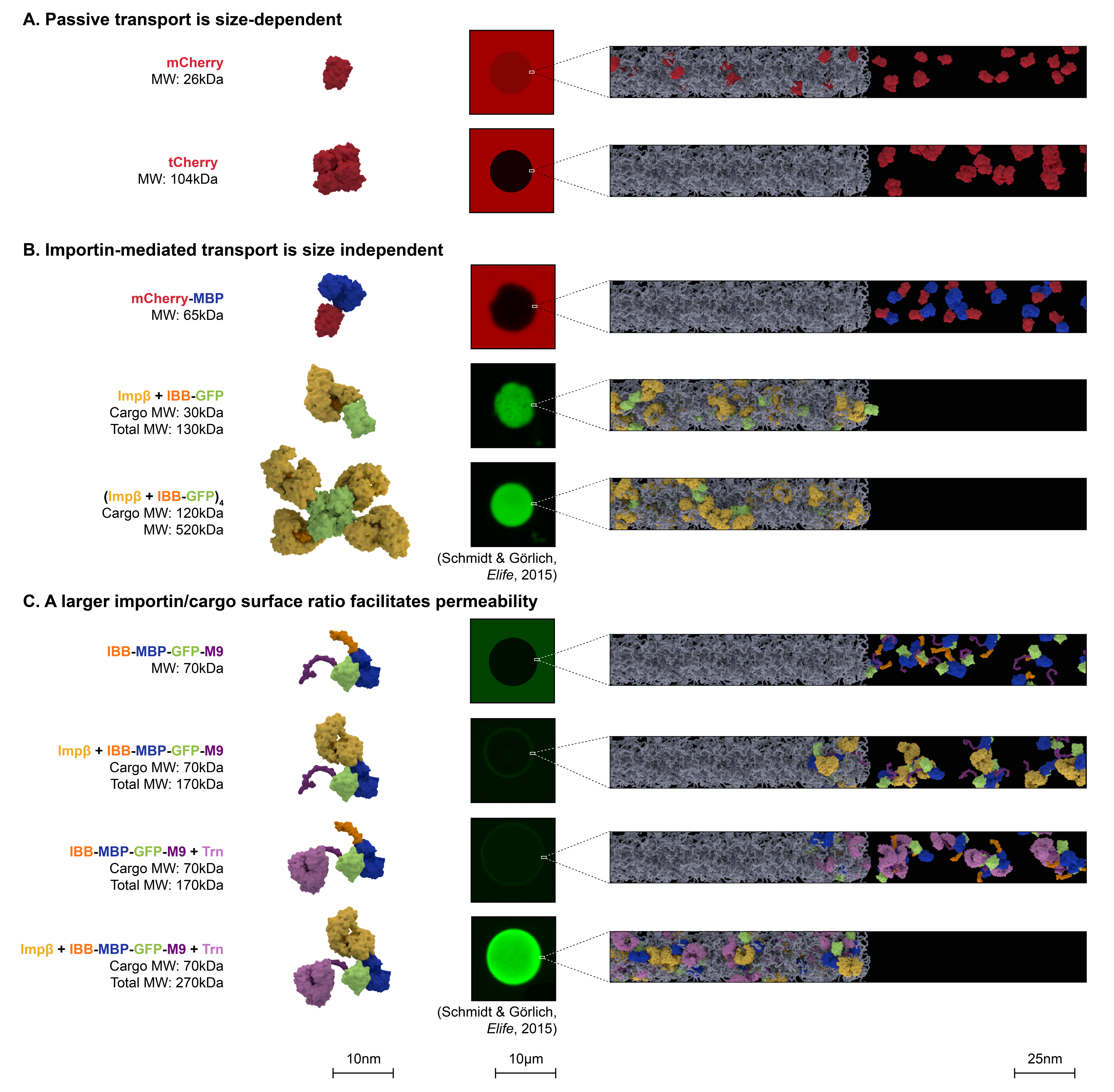
Droplets made of FG-Nups, like the ones shown above, were able to recapitulate the salient features of nuclear transport: free diffusion of small molecules, such as mCherry (MW = 26kD), and exclusion of larger molecules, such as tetrameric Cherry (tCherry, MW = 104kD).

Large molecules, however, were able to enter the droplet if importins were included in the reaction, such as the tetrameric fluorophore zsGreen bound to four importins (MW = 520kD).

Importins were more efficient in allowing diffusion into the droplets when the surface ratio between importins and their cargo was higher. For example, MBP-GFP (MW = 70kD) only entered the edges of the droplet when interacting with one importin, but diffused deep into the droplet when interacting with two importins. This is consistent with the idea that importins and exportins locally disassemble the barrier sieve.

The droplets also recreated the concentrating feature of nuclear transport. Small molecules, like mCherry, diffused into the droplets until their concentration was roughly equal inside and outside the droplet. Importin-cargo complexes, however, not only entered the droplets, but were concentrated in them, reaching concentrations higher than in the surrounding regions.

3. A concrete example: nucleocytoplasmic transport plays a key role in the regulation of NF-kB signalling

NF-κB is an ubiquitous transcription factor fundamental in the innate immune response. Upon stimulation by a range of external signals such as TNFα or other inflammatory cytokines, it exhibits oscillatory nucleocytoplasmic shuttling. This alternating subcellular localization translates into oscillatory activation of downstream signaling pathways, and thereby transmits information about the sequence of external signal pulses.
Constitutive NF-κB activation is a hallmark of most cancers, where its activity sustains cell survival and proliferation, among many other aspects of cancer biology. Some of the therapeutic approaches that are currently being examined are perturbations to nuclear import export as means to downregulate NF-κB. See Verzella & al (Biomedicines, 2022), Nachmias & Schimmer (Leukemia, 2020), or Gilmore (Biomedicines, 2021) for more information about NF-κB and cancer.

The following animation highlights the crucial regulatory role played by nucleocytoplasmic transport in the NF-κB–IκB feedback loop, a canonical example of transcription factor signaling.