Illustrating Necroptosis
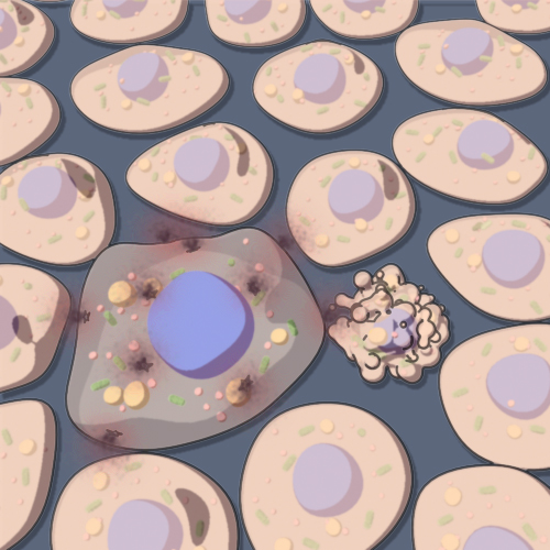
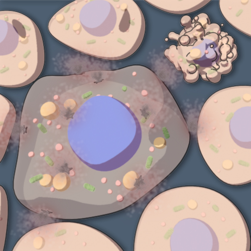
In contrast to apoptosis, which is defined as a type of programmed cell death, necrosis has long been thought to represent an unregulated form of cell death which is triggered by cellular trauma or stress. Recent studies have shown, however, that some forms of necrosis are regulated using a molecular pathway that is partially intertwined with that of the apoptotic regulatory network. This process has been termed necroptosis.
The goal of this project was to create an illustration that compares the necroptotic cell phenotype to that of an apoptotic cell. During necroptosis, the major distinguishing features are a significant increase in the size of the cell, and a greater transparency of the cell that is likely due to the entry of water and expulsion of cytoplasmic material through perforations in the plasma membrane. In addition, the nucleus appears larger in the necrotic cell, and there is a small increase in vesicles and an increase in lysosomes size. In comparison, apoptotic cells appear smaller, with a large amount of blebbing of the plasma membrane and swelling of the mitochondria.


Animating the necroptosis pathway
There is an increasing amount of experimental evidence that shows that the same signaling pathways that mediate apoptotic cell death also mediate death by necroptosis. The animation shown below is an exploration of the signaling pathway leading to necroptotic cell death.
Acknowledgements
Many thanks to Junying Yuan and Dana Christofferson (Harvard Medical School) for collaborating on this project.


